
Background:
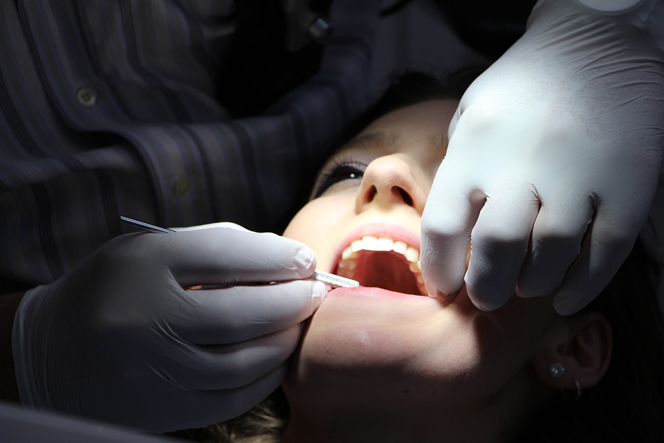
As children we are told to brush our teeth and floss twice a day. An uncomfortable dental experience as a child can create dental anxiety in adulthood. This anxiety can prevent patients from seeking the care they need. The development of caries, or cavities, in the mouth can have lasting impacts if not treated properly in both children and adults. Caries develop on teeth due to the creation of an acidic environment from the bacteria inhabiting the mouth (Marsh 1994). The acidity eats away at the enamel, and eventually the softer pulp, or dentin, within the tooth, leaving an exposed and sensitive area that requires medical attention (Bowen et al. 2017). The prevalence of decay in primary teeth is notably higher in children aged 1 to 4 while young adults aged 15-19 years old have a higher prevalence of decay present in permanent teeth (Kassebaum et al. 2017). Because children develop higher levels of cavities than adults, protecting young permanent teeth is an important area for research. Why are children more likely to develop caries? And what can be done to lessen the spread of this disease? For many years scientists attributed the development of caries to the bacterial species Streptococcus mutans. With the advent of new technology and further information of how microbes interact with each other, scientists are now looking at old problems with modern solutions. Instead of merely describing the presence of microbes, scientists are now beginning to see what is happening between various microbial species in the human microbiome.
Central Goal:
In a recent study, researchers aimed to characterize the microbiome of dental plaque in twins with and without dental caries. Additionally, researchers categorized specific genes and pathways the microbial community could be using in the diseased and non-diseased state. (Espinoza et al. 2018).
Evidence:
After sequencing the DNA from the twin research participants, Espinoza et al. (2018) were able to begin to categorize and compare the taxa present in the sample. Unsurprisingly, researchers found 119 Streptococcus species including the villainous S. mutans. Analysis indicated a higher level of microbial diversity in the healthy communities without caries. Even though communities associated with caries had a lower level of microbial diversity, there was a higher level of Streptococcus diversity.
Espinoza et al. (2018) concluded that caries can be detected by looking for microbes other than Streptococcus mutans as there is a common core group of species between all of the diseased samples. Taxa such as Alloprevotella and Gracilibacteria were also discovered. Both taxa may contribute to the formation of cavities through glucose fermentation and the production of acids. Healthy samples had lower levels of Streptococcus diversity which can also serve as a biomarker for the development of decay. This idea challenges the common thought that Streptococcus mutans can cause caries, but is backed up by several other ecological studies. The researchers posited the change in how sugar is metabolized corresponds with a change in the microbes present and the creation of acidic products which cause decay. An environment can move from healthy to diseased by introducing different types of sugars which the microbes break down into acidic products and lower the pH. As the pH lowers, more caries-causing microbes are selected for and the pH continues to decrease, lowering the diversity of the diseased environment. This positive feedback system is similar to a rolling a snowball. As the size of the snowball increases, more snow can be added, increasing the surface area. Eventually, a fist-sized ball grows to a diameter of feet, not inches. This phenomena was seen in the difference in microbial diversity between the healthy and diseased state. Additionally the presence of specific proteins in the diseased samples which respond to environmental stimuli could enable the caries-causing bacteria to be able to withstand lower pH levels.
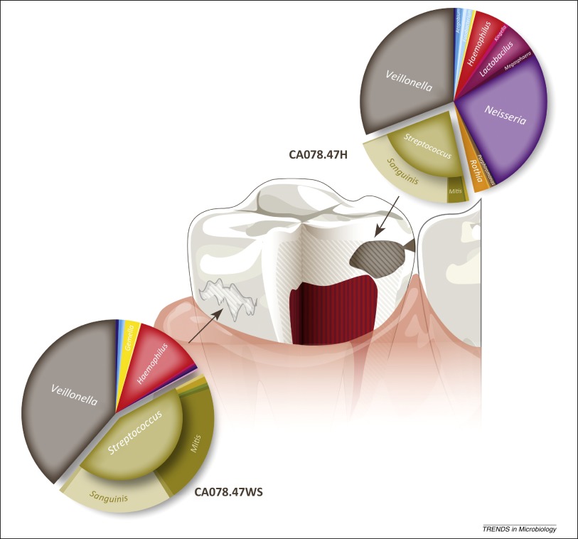
After discovering what microbes were present in the plaque biofilm found on the crowns of teeth, the next step was to see what those microbes were doing. Additionally, they delved even further to see if the presence of these microbes could be traced back to the development of dental caries in children. After sequencing all of the DNA present in the samples, the researchers were able to match the DNA sequences with already discovered genetic pathways. Differences in the DNA sequences found can help to distinguish different taxa and determine what pathways they provide to the community. In the caries-causing communities, they found higher abundance of various sugar uptake pathways and specific signaling patterns from their environment. Researchers also identified the presence of antibiotic resistance strategies, including tetracycline and bacitracin resistance, and genes related to metal transport. Grouping the DNA of the taxa together enabled the researchers to create 11 metagenome assembled genomes to further compare the different taxa discovered in the samples. By comparing and contrasting the different taxa, researchers can determine what genes are present in the communities and determine how they work together to produce the diseased state, in this instance, dental caries. By comparing the genes which were sequenced, two important taxa were identified including the lesser known candidate taxa TM7 (Saccharibacteria) and the newly discovered Gracilibacteria which may live inside other microbes to the benefit of both.
Breaking down enamel and dentin releases toxic levels of metal ions. Caries-causing microbes require the presence of metal transport ions to minimize the toxicity of the ions. The disease causing microbes are able to proliferate in these areas because they have the cellular machinery to remove the metals from their environment. Researchers were not able to determine if the taxa present used any of the genes they sequenced and tie function to taxa because not all taxa use every gene present in their genome. They were able to describe various niches in the community and further concluded the whole community is responsible for causing caries. While a core microbiome was found between all the samples of twin children, the outliers are responsible for creating a healthy or diseased state. The presence of various sugars can change the entire community from a healthy to a diseased state.
My Questions:
Now that we know how the oral microbiome changes and caries occur, more research needs to be done to see if these processes can be prevented. Are certain types of sugar more likely to cause the changes noted in the paper over others? Perhaps the kind of sugar we eat matters more than the amount of sugar. Are there other foods that contribute to a diseased state? This paper has led researchers to begin questioning what genetic pathways are really being used in this environment and if any differences matter. More knowledge on this subject could lead to better prevention strategies and more effective treatment therapies for children and adults alike.
Further Reading:
Interested in more about dental caries research? Check out these papers!
- Host Genetic Control of the Oral Microbiome in Health and Disease Gomez et. al (2017) discovered that while the environment plays a large role in the oral microbiome, heritable oral taxa have also been found. These taxa were not attributed to caries and their abundance decreased with age and lowered sugar intake. While genetics are important, they do not seem to contribute to the development of caries.
- A Novel Antimicrobial Peptide Against Dental-Caries-Associated Bacteria Chen et al. (2017) recognized the potential effectiveness of the antimicrobial peptide ZXR-2 to disrupt biofilm formation in S. mutans and other gram-negative and gram-positive bacteria. This treatment could be a potential therapy for caries prevention and treatment.
- Identifying a Healthy Oral Microbiome through Metagenomics Alcaraz et al. (2012) applied a metagenomics approach to describing the oral microbiome. They found similar results to using 16S rRNA amplification which allows researchers to use this new method to describe the genetic make-up of microbial communities in the mouth.
- Quantitative Analysis of Biofilm Bacteria According to Different Stages of Early Childhood Caries The formation of biofilms plays a major role in the development of dental caries. Gonçalves Neves et al. (2018) identified a strong association between the presence of Streptococcus mutans and Bifidobacterium spp. in biofilms and the development of caries in Brazilian children with various levels of dental caries.
- The Structure of Dental Plaque Microbial Communities in the Transition from Health to Dental Caries and Periodontal Disease Understanding what dental plaque is made out of is essential to preventing its growth in the first place. Valm (2019) condenses known information regarding how an imbalance of a healthy oral microbiome can lead to the formation of dental caries. Valm highlights the changes in healthy biofilms to disease causing biofilms.
References:
- Bowen, W.H., Burne, R.A., Wu, H., Koo, H. 2017. Oral biofilms: pathogens, matrix, and polymicrobial interactions in microenvironments. Trends in Microbiology 26(3):229-242. Doi: 10.1016/j.tim.2017.09.008
- Espinoza, J.L., Harkins, D.M., Torralba, M., Gomez, A., Highlander, S.K., Jones, M.B., Leong, P., Saffery, R., Bockman, M., Kuelbs, C., Inman, J.M., Hughes, T., Craig, J.M., Nelson, K.E., Dupont, C.L. 2018. Supragingival plaque microbiome ecology and functional potential in the context of health and disease. mBio 9:e01631-18. Doi: 10.1128/mBio.01631-18
- Kassebaum, N.J., Smith, A.G.C., Bernabé, E., Fleming, T.D., Reynolds, A.E., Vos, T., Murray, C.J.L., Marcenes, W., & GBD 2015 Collaborators. 2017. Global, regional, and national prevalence, incidence, and disability-adjusted life years for oral conditions for 195 countries, 1990-2015: A systematic analysis for the global burden of diseases, injuries, and risk factors. Journal of Dental Research 96(4):380-387. Doi: 10.1177/0022034517693566
- Marsh, P.D. 1994. Microbial ecology of dental plaque and its significance in health and disease. Advances in Dental Research 8(2):263-271. Doi: 10.1177/08959374940080022001
- Simón-Soro, A. & Mira, A. 2015. Solving the etiology of dental caries. Trends in Microbiology 23(2):76-82. Doi: 10.1016/j.tim.2014.10.010